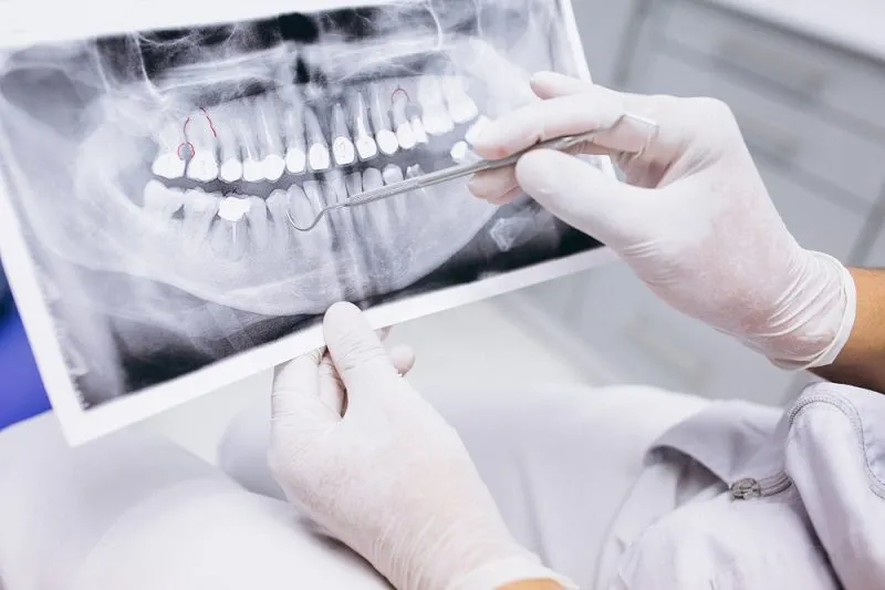
What Are Dental X-Rays?
Dental X-rays are diagnostic imaging examinations that use low-dose X-ray radiation to evaluate the condition of teeth, jawbone, tooth roots, and surrounding tissues that cannot be fully assessed through a standard clinical examination alone.
Through dental radiographs, dentists can detect hidden issues such as cavities between teeth, root infections, impacted wisdom teeth, and jawbone conditions before performing certain treatments.
Today, panoramic dental X-rays and modern digital imaging technologies allow examinations to be faster, more comfortable, and more accurate while exposing patients to significantly lower radiation levels compared to older technologies.
At Onyx Dental Center, radiographic examinations play an important role in helping dentists make more precise diagnoses before treatments such as dental implants, orthodontics, or root canal therapy.
Types of Dental X-Rays
Periapical X-Ray
A periapical X-ray is used to examine one or several teeth in detail, from the crown to the tip of the root.
This type of X-ray is commonly used to:
Detect infections at the root tip
Evaluate deep cavities
Assist during root canal treatment (RCT)
Assess surrounding bone conditions
Because it provides highly detailed images, this X-ray is often recommended when patients experience pain in a specific tooth.
Panoramic X-Ray
A panoramic dental X-ray captures the entire upper and lower jaw in a single image.
This examination helps dentists evaluate:
Wisdom tooth position
Impacted teeth
Jaw abnormalities
Tooth number and positioning
Orthodontic and implant treatment planning
At Onyx Dental Center, panoramic X-rays are frequently used as an initial examination for comprehensive oral evaluations.
Bitewing X-Ray
Bitewing X-rays are used to view the crowns of upper and lower teeth simultaneously, especially the spaces between teeth.
This type is highly effective for detecting:
Cavities between teeth
Leaking dental fillings
Mild tartar buildup below the gumline
Because many cavities begin between teeth where they are difficult to detect visually, bitewing X-rays are an important part of routine dental check-ups.
CBCT (Cone Beam Computed Tomography)
CBCT is a 3D dental imaging technology that provides highly detailed views of the teeth and jawbone.
CBCT is commonly used for:
Dental implant planning
Evaluation of complex impacted teeth
Jawbone analysis
Oral surgery cases
Complex root canal evaluations
This technology allows dentists to visualize nerve pathways, bone thickness, and jaw anatomy more accurately than conventional X-rays.
What Can Dental X-Rays Detect?
Many dental problems cannot be seen directly during routine examinations or when patients look in the mirror.
Dental X-rays can help detect:
Hidden cavities
Root infections
Dental abscesses
Cysts or jawbone abnormalities
Impacted wisdom teeth
Bone infections caused by gum disease
Tooth positioning before orthodontic treatment
Bone conditions before implant placement
Dental radiographs often become an essential part of diagnosis and treatment planning.
When Do Dentists Recommend Dental X-Rays?
Not every patient requires X-rays at every visit. Dentists usually consider clinical findings, dental history, age, and oral disease risk before recommending imaging.
Dental X-rays may be recommended for:
Tooth pain with unclear causes
Suspected infection or abscess
Impacted wisdom teeth
Orthodontic or clear aligner planning
Before dental implant placement
Gum disease evaluation
Dental trauma or injury
Routine check-ups in high-caries-risk patients
Dentists only recommend dental X-rays when they are necessary to support diagnosis, treatment success, and early problem detection.
Are Dental X-Rays Safe? Understanding Radiation Exposure
One of the most common concerns is whether dental X-rays are safe.
In general, modern dental X-rays use very low doses of radiation and are considered safe when performed according to medical indications.
Digital radiography technology has significantly reduced radiation exposure compared to older conventional systems.
Dentists also follow the ALARA principle (As Low As Reasonably Achievable), meaning radiation exposure is kept as low as possible while still obtaining the information needed for accurate diagnosis.
Patients therefore do not need to be overly worried about medically necessary dental X-rays.
Comparing Dental X-Ray Radiation to Everyday Exposure
Radiation exposure from dental X-rays is relatively small compared to natural daily background radiation.
For example:
Single Tooth X-Rays (Bitewing / Periapical)
These small X-rays focus on only a few teeth. Four bitewing or periapical X-rays generally expose patients to radiation equivalent to approximately one day of natural environmental radiation.
Panoramic X-Rays
Although panoramic images cover the entire jaw, the radiation dose remains very low and is considered safe.
3D CBCT Imaging
CBCT uses slightly higher radiation because it creates detailed 3D images. However, the dose is still tightly regulated to remain within safe medical standards.
Modern dental imaging systems are also designed to focus radiation only on the area being examined, minimizing unnecessary exposure.
Radiation exposure is also naturally encountered from:
Sunlight
Air travel
Natural environmental sources
Because of this, the diagnostic benefits of dental X-rays generally far outweigh the minimal risks.
Can Pregnant Women and Children Have Dental X-Rays?
Pregnant Women
Many pregnant women worry that dental X-rays may affect the baby. In reality, dental X-rays are generally considered safe during pregnancy when medically necessary.
Important facts include:
Dental X-ray beams are highly focused on the mouth and jaw area, not the abdomen or uterus.
Radiation exposure to the fetus is extremely minimal—almost negligible according to radiation safety guidelines.
Dentists provide additional protection using lead aprons over the abdomen during the procedure.
In fact, delaying treatment for severe dental infections due to fear of X-rays may pose greater health risks to both mother and baby.
Important Tip: Always inform your dentist if you are pregnant or planning pregnancy. Dentists will choose the safest timing and lowest-radiation imaging methods whenever possible.
Children
Dental X-rays may also be necessary for children to:
Monitor tooth development
Evaluate permanent teeth growth
Assess tooth positioning
Detect hidden cavities
Radiation doses are carefully adjusted according to the child’s age and clinical needs.
How Often Should Dental X-Rays Be Taken?
There is no universal rule that everyone must undergo dental X-rays every few months.
The frequency depends on:
Risk of cavities
Oral health condition
History of dental disease
Age
Type of treatment being performed
Patients with high cavity risk or gum disease may require more frequent imaging compared to individuals with healthy oral conditions.
Dentists determine imaging needs individually for each patient.
Conclusion
Dental X-rays are important diagnostic tools that help dentists detect problems not visible during routine oral examinations. Technologies such as panoramic X-rays and CBCT enable more accurate diagnoses and safer, more effective treatment planning.
Radiation exposure from modern dental X-rays is low and generally considered safe when performed according to proper medical indications. Patients therefore should not hesitate to undergo imaging when recommended by their dentist.
If you experience tooth pain, jaw discomfort, or would like a comprehensive oral evaluation, do not delay consulting your dentist before problems become more serious.
References
Lubis, L. E., Bayuadi, I., Bayhaqi, Y. A., Ardiansyah, F., Setiadi, A. R., Sugandi, R. D., Craig, L. A., Nasir, A., Basith, R. A., Pawiro, S. A., & Soejoko, D. S. (2019). RADIATION DOSE from DENTAL RADIOGRAPHY in INDONESIA: A FIVE-YEAR SURVEY. Radiation Protection Dosimetry, 183(3), 342-347. https://doi.org/10.1093/rpd/ncy123
Rahmi-Fajrin, H., Puspita, S., Riyadi, S., & Sofiani, E. (2018). Dental radiography image enhancement for treatment evaluation through digital image processing. Journal of clinical and experimental dentistry, 10(7), e629–e634. https://doi.org/10.4317/jced.54607
Schüler, I. M., Hennig, C. L., Buschek, R., Scherbaum, R., Jacobs, C., Scheithauer, M., & Mentzel, H. J. (2023). Radiation Exposure and Frequency of Dental, Bitewing and Occlusal Radiographs in Children and Adolescents. Journal of personalized medicine, 13(4), 692. https://doi.org/10.3390/jpm13040692